Promising Therapy for Cardiac Regeneration
New research gives information in order to understand safety, efficacy, and mechanisms of action of a new cardiac therapy.
Ischemic heart disease (IHD) has maintained its rank as one of the worldwide leading causes of mortality outweighing the burden from all malignancies combined.
When IHD develops, chronic myocardial ischemia, aggravated in some instances by periods of acute ischemia in the form of myocardial infarction, ensue. Damaged myocardium is replaced with a fibrotic scar that over-activates physiologic compensatory mechanisms with challenging sequalae, such as myocardial rigidity and eventually, over time, heart failure.
A research collaboration team at University of Helsinki together with a State Key Laboratory of Cardiovascular Disease (FuWai Hospital, Beijing, China) has investigated in a mouse model of artificial myocardial infarction, the molecular mechanisms underlying novel, easily clinically implementable tissue-engineered approach for stimulating the myocardial regeneration.
The tissue-engineered approach relies on a local transplantation of minute pieces of autologous atrial appendage tissue, termed atrial appendage micrografts (AAMs), to the surface of the ischaemically stressed myocardium.
Results of the investigation are published in the Journal of Heart and Lung Transplantation.
"We were able to get a comprehensive view on how the heart's functional, structural and metabolic aspects of healing are influenced by AAMs patch transplantation following acute ischemia," says Docent Esko Kankuri from University of Helsinki.
Heart's pumping function preserved, also improved functional recovery
The research group utilized complementary tools, including systematic postoperative functional echocardiographic follow-up, histomorphometric analyses and finally site-selective proteomics in tandem with functional bioinformatics.
"We demonstrated not only preservation of heart's pumping function following critical ischemic insult but also improved functional recovery following AAMs transplantation during follow-up," says Docent Maciej Lalowski from University of Helsinki.
"We identified 1005 proteins from the myocardium, of which 216 were differentially expressed immediately below the AAMs patch in the 'subtransplant' area and 43 in the interventricular septum remote to the AAMs transplantation site," Lalowski continues.
The therapy is currently undergoing clinical safety and feasibility evaluation as an adjuvant to the coronary artery bypass grafting operation.
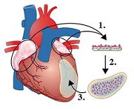
Illustration: Schematic overview of the atrial appendage micrografts (AAMs) patch transplantation. 1: The material is collected. 2: Setup with extracellular matrix (ECM) patch. 3: The AAMs patch is transplanted epicardially on the injured site.
Read more...
University of Helsinki News Release (06/30/20)
Science Daily (06/30/20)
Abstract (The Journal of Heart and Lung Transplantation, 2020.)
When IHD develops, chronic myocardial ischemia, aggravated in some instances by periods of acute ischemia in the form of myocardial infarction, ensue. Damaged myocardium is replaced with a fibrotic scar that over-activates physiologic compensatory mechanisms with challenging sequalae, such as myocardial rigidity and eventually, over time, heart failure.
A research collaboration team at University of Helsinki together with a State Key Laboratory of Cardiovascular Disease (FuWai Hospital, Beijing, China) has investigated in a mouse model of artificial myocardial infarction, the molecular mechanisms underlying novel, easily clinically implementable tissue-engineered approach for stimulating the myocardial regeneration.
The tissue-engineered approach relies on a local transplantation of minute pieces of autologous atrial appendage tissue, termed atrial appendage micrografts (AAMs), to the surface of the ischaemically stressed myocardium.
Results of the investigation are published in the Journal of Heart and Lung Transplantation.
"We were able to get a comprehensive view on how the heart's functional, structural and metabolic aspects of healing are influenced by AAMs patch transplantation following acute ischemia," says Docent Esko Kankuri from University of Helsinki.
Heart's pumping function preserved, also improved functional recovery
The research group utilized complementary tools, including systematic postoperative functional echocardiographic follow-up, histomorphometric analyses and finally site-selective proteomics in tandem with functional bioinformatics.
"We demonstrated not only preservation of heart's pumping function following critical ischemic insult but also improved functional recovery following AAMs transplantation during follow-up," says Docent Maciej Lalowski from University of Helsinki.
"We identified 1005 proteins from the myocardium, of which 216 were differentially expressed immediately below the AAMs patch in the 'subtransplant' area and 43 in the interventricular septum remote to the AAMs transplantation site," Lalowski continues.
The therapy is currently undergoing clinical safety and feasibility evaluation as an adjuvant to the coronary artery bypass grafting operation.
Illustration: Schematic overview of the atrial appendage micrografts (AAMs) patch transplantation. 1: The material is collected. 2: Setup with extracellular matrix (ECM) patch. 3: The AAMs patch is transplanted epicardially on the injured site.
Read more...
University of Helsinki News Release (06/30/20)
Science Daily (06/30/20)
Abstract (The Journal of Heart and Lung Transplantation, 2020.)